Leg Bone Diagram / Foot Bones Anatomy Conditions And More
Leg Bone Diagram / Foot Bones Anatomy Conditions And More. Some types of leg pain can be traced to problems in your lower spine. The knee joint is the largest joint in the body and is primarily a hinge joint, although some sliding and rotation occur. With different grades of sprains depending on severity. These muscles work together to produce movements such as standing, walking, running, and jumping. The femur, or thighbone, is the longest and largest bone in the human body.
ads/bitcoin1.txt
The major bones of the leg are the femur (thigh bone), tibia (shin bone), and adjacent fibula, and these are all long bones.the patella (kneecap) is the sesamoid bone in front of the knee.most of the leg skeleton has bony prominences and margins that can be palpated and some serve as anatomical landmarks that define the extent of the leg. The quadriceps muscle attachment points. The lower leg extends from the knee to the ankle. There are two types of cartilaginous joints: Also called the shin bone, the tibia is the longer of the two bones in the.
Scanning X Ray Image Of Lower Leg Bone Download Scientific Diagram from www.researchgate.net The femur, or thighbone, is the longest and largest bone in the human body. Look at links below to get more options for getting and using clip art. The pubis, ischium, and ilium together constitute the pelvis while the thigh bone is the femur. See more ideas about muscle anatomy, human anatomy and physiology, body anatomy. Most leg pain results from wear and tear, overuse, or injuries in joints or bones or in muscles, ligaments, tendons or other soft tissues. The lower leg extends from the knee to the ankle. There are two types of cartilaginous joints: Learn vocabulary, terms, and more with flashcards, games, and other study tools.
The hip itself is a ball and socket joint, much like the shoulder.the structures necessary to create this joint are the socket, the joint capsule, muscle, ligaments, and the neck.
ads/bitcoin2.txt
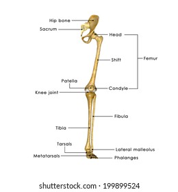
It also separates muscles on the anterior and posterior parts of the leg. There are two types of cartilaginous joints: The diagram of bones in the ankle and foot is given below: The femur, or thigh bone, is the single bone of the thigh region (figure 6.51). The bones of the hip include the femur, the ilium, the ischium, and the pubis. These muscles work together to produce movements such as standing, walking, running, and jumping. Now let's look at the tibia bone, which is the larger of the two leg bones, located medially. Pelvic bone labeled 12 photos of the pelvic bone labeled pelvic bone labeled, pelvic bone labeling quiz. Leg bone anatomy diagram diagram of human leg human anatomy diagram 10 / 10 ( 1 vote ) in this image, you will find femur, medial epicondyle of the femur, patella, tibial tuberosity, anterior rest of the tibia, a medial surface of the tibia, lateral epicondyle of the femur, head of the fibula, fibula, medial malleolus of the tibia, lateral. The lower leg is comprised of two bones, the tibia and the smaller fibula. The femur, or thighbone, is the longest and largest bone in the human body. Learn vocabulary, terms, and more with flashcards, games, and other study tools. With different grades of sprains depending on severity.
By tightening and relaxing, the skeletal muscles create movement. The bones of the leg and foot form part of the appendicular skeleton that supports the many muscles of the lower limbs. The tarsal bones in the foot are located amongst tibia, metatarsal bones, and fibula. A long bone is a bone that has greater length than width. With different grades of sprains depending on severity.
Bones Of The Leg And Foot Interactive Anatomy Guide from www.innerbody.com They support bones, in this case, the vertebrae. Long bones have a thick outside layer of compact bone and an inner medullary cavity containing bone marrow. With different grades of sprains depending on severity. Most leg pain results from wear and tear, overuse, or injuries in joints or bones or in muscles, ligaments, tendons or other soft tissues. There are in all 7 bones, which fall under tarsal bones category. The knee joint is the largest joint in the body and is primarily a hinge joint, although some sliding and rotation occur. The diagram of bones in the ankle and foot is given below: The thigh bone, or femur, is the large upper leg bone that connects the lower leg bones (knee joint) to the pelvic bone (hip joint).
Related posts of diagram of leg bones pelvic bone labeled.
ads/bitcoin2.txt
The tibia, commonly known as the 'shin bone', is the largest and most medial of the two.you can palpate its anterior border when you run your finger down the anterior aspect of your leg. The lower leg is comprised of two bones, the tibia and the smaller fibula. The bones of the leg are the femur, tibia, fibula and patella.the foot bones shown in this diagram are the talus, navicular, cuneiform, cuboid, metatarsals and calcaneus. The knee joint is the largest joint in the body and is primarily a hinge joint, although some sliding and rotation occur. The hip itself is a ball and socket joint, much like the shoulder.the structures necessary to create this joint are the socket, the joint capsule, muscle, ligaments, and the neck. Structure of anatomy leg and foot 6 photos of the structure of anatomy leg and foot leg foot anatomy, leg foot bones, leg foot cramps, leg foot cramps at night, leg foot massage, leg foot numbness, leg foot pain, leg foot tattoos, foot, leg foot anatomy, leg foot. The back muscles are skeletal muscles. See more ideas about muscle anatomy, human anatomy and physiology, body anatomy. The quadriceps muscle attachment points. There are two types of cartilaginous joints: It also separates muscles on the anterior and posterior parts of the leg. Pelvic bone labeled 12 photos of the pelvic bone labeled pelvic bone labeled, pelvic bone labeling quiz. The tarsal bones in the foot are located amongst tibia, metatarsal bones, and fibula.
Most leg pain results from wear and tear, overuse, or injuries in joints or bones or in muscles, ligaments, tendons or other soft tissues. A long bone is a bone that has greater length than width. By tightening and relaxing, the skeletal muscles create movement. Some types of leg pain can be traced to problems in your lower spine. Structure of anatomy leg and foot 6 photos of the structure of anatomy leg and foot leg foot anatomy, leg foot bones, leg foot cramps, leg foot cramps at night, leg foot massage, leg foot numbness, leg foot pain, leg foot tattoos, foot, leg foot anatomy, leg foot.
Labelled Bones Leg Images Stock Photos Vectors Shutterstock from image.shutterstock.com The bones of the hip include the femur, the ilium, the ischium, and the pubis. Our goal is that these leg anatomy worksheets pictures gallery can be a direction for you, bring you more references and also make you have a great day. Start studying lab test 2: The quadriceps muscle attachment points. Pelvic bone labeled 12 photos of the pelvic bone labeled pelvic bone labeled, pelvic bone labeling quiz. Leg pain can also be caused by blood clots, varicose veins or poor circulation. A long bone has a shaft and 2 ends. *the origin, insertion, and belly.* a muscle's origin is where a tendon attaches it to the *less* movable bone.
The knee joint is the largest joint in the body and is primarily a hinge joint, although some sliding and rotation occur.
ads/bitcoin2.txt
The bones of the leg are the femur, tibia, fibula and patella.the foot bones shown in this diagram are the talus, navicular, cuneiform, cuboid, metatarsals and calcaneus. It also separates muscles on the anterior and posterior parts of the leg. All four quadriceps muscles insert into the tibia (shin bone). The tarsal bones in the foot are located amongst tibia, metatarsal bones, and fibula. *the origin, insertion, and belly.* a muscle's origin is where a tendon attaches it to the *less* movable bone. Leg pain can also be caused by blood clots, varicose veins or poor circulation. The bones of the hip include the femur, the ilium, the ischium, and the pubis. There are two types of cartilaginous joints: At the same time, the bones and joints of the leg and foot must be strong enough to support the body's weight while remaining. Every skeletal muscle has three main parts: The major bones of the leg are the femur (thigh bone), tibia (shin bone), and adjacent fibula, and these are all long bones.the patella (kneecap) is the sesamoid bone in front of the knee.most of the leg skeleton has bony prominences and margins that can be palpated and some serve as anatomical landmarks that define the extent of the leg. Related posts of diagram of leg bones pelvic bone labeled. The back muscles are skeletal muscles.
ads/bitcoin3.txt
ads/bitcoin4.txt
ads/bitcoin5.txt
0 Response to "Leg Bone Diagram / Foot Bones Anatomy Conditions And More"


0 Response to "Leg Bone Diagram / Foot Bones Anatomy Conditions And More"
Post a Comment